Hello
How may I help you today?
-
24/7 available services
-
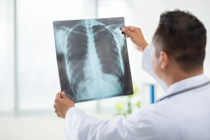
X-rays use invisible electromagnetic energy beams to produce images of internal tissues, bones, and organs on film or digital media. Standard X-rays are performed for many reasons, including diagnosing tumors or bone injuries.
X-rays are made by using external radiation to produce images of the body, its organs, and other internal structures for diagnostic purposes. X-rays pass through body structures onto specially-treated plates (similar to camera film) or digital media and a "negative" type picture is made (the more solid a structure is, the whiter it appears on the film).
When the body undergoes X-rays, different parts of the body allow varying amounts of the X-ray beams to pass through. .
What is a CT scan?
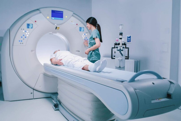
A CT (computed tomography) scan is a type of imaging test. Like an X-ray, it shows structures inside your body. But instead of creating a flat, 2D image, a CT scan takes dozens to hundreds of images of your body. To get these images, a CT machine takes X-ray pictures as it revolves around you.
Healthcare providers use CT scans to see things that regular X-rays can't show. For example, body structures overlap on regular X-rays and many things aren't visible. A CT shows the details of each of your organs for a clearer and more precise view.
Another term for CT scan is CAT scan. CT stands for "computed tomography," while CAT stands for "computed axial tomography." But these two terms describe the same imaging test.
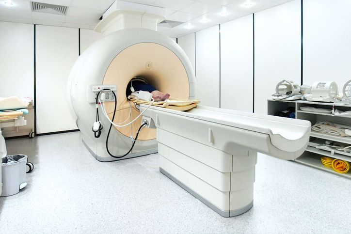
The MRI machine is a large, cylindrical (tube-shaped) machine that creates a strong magnetic field around the patient and sends pulses of radio waves from a scanner. Some MRI machines look like narrow tunnels, while others are more open.
The strong magnetic field created by the MRI scanner causes the atoms in your body to align in the same direction. Radio waves are then sent from the MRI machine and move these atoms out of the original position. As the radio waves are turned off, the atoms return to their original position and send back radio signals. These signals are received by a computer and converted into an image of the part of the body being examined. This image appears on a viewing monitor.
MRI may be used instead of computed tomography (CT) when organs or soft tissue are being studied. MRI is better at telling the difference between types of soft tissues and between normal and abnormal soft tissues.
An echocardiogram is a noninvasive (the skin is not pierced) procedure used to assess the heart's function and structures. During the procedure, a transducer (like a microphone) sends out sound waves at a frequency too high to be heard. When the transducer is placed on the chest at certain locations and angles, the sound waves move through the skin and other body tissues to the heart tissues, where the waves bounce or "echo" off of the heart structures. These sound waves are sent to a computer that can create moving images of the heart walls and valves.
An echocardiogram may use several special types of echocardiography, as listed below:
M-mode echocardiography. This, the simplest type of echocardiography, produces an image that is similar to a tracing rather than an actual picture of heart structures. M-mode echo is useful for measuring or viewing heart structures, such as the heart's pumping chambers, the size of the heart itself, and the thickness of the heart walls.
Doppler echocardiography. This Doppler technique is used to measure and assess the flow of blood through the heart's chambers and valves. The amount of blood pumped out with each beat is an indication of the heart's functioning. Also, Doppler can detect abnormal blood flow within the heart, which can indicate a problem with one or more of the heart's four valves, or with the heart's walls.
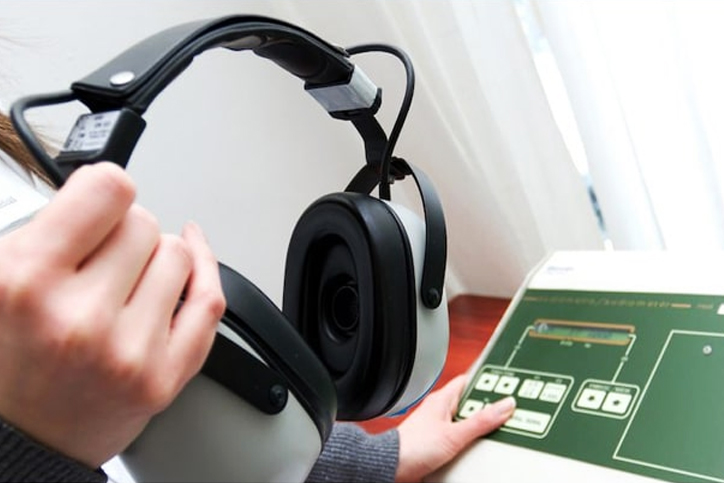
Audiometry consists of tests of function of the hearing mechanism. This includes tests of mechanical sound transmission (middle ear function), neural sound transmission (cochlear function), and speech discrimination ability (central integration). A complete evaluation of a patient's hearing must be done by trained personnel using instruments designed specifically for this purpose.
Pure tones (single frequencies) are used to test air and bone conduction. These and speech testing are done with an audiometer. The audiometer is an electric instrument consisting of a pure tone generator, a bone conduction oscillator for measuring cochlear function, an attenuator for varying loudness, a microphone for speech testing, and earphones for air conduction testing.
Other tests include impedance audiometry, which measures the mobility and air pressure of the middle ear system and middle ear (stapedial) reflexes, and auditory brainstem response (ABR), which measures neural transmission time from the cochlea through the brainstem.
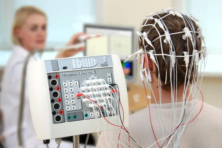
An EEG is a test that detects abnormalities in your brain waves, or in the electrical activity of your brain. During the procedure, electrodes consisting of small metal discs with thin wires are pasted onto your scalp. The electrodes detect tiny electrical charges that result from the activity of your brain cells. The charges are amplified and appear as a graph on a computer screen, or as a recording that may be printed out on paper. Your healthcare provider then interprets the reading.
During an EEG, your healthcare provider typically evaluates about 100 pages, or computer screens, of activity. He or she pays special attention to the basic waveform, but also examines brief bursts of energy and responses to stimuli, such as flashing lights.
Evoked potential studies are related procedures that also may be done. These studies measure electrical activity in your brain in response to stimulation of sight, sound, or touch.
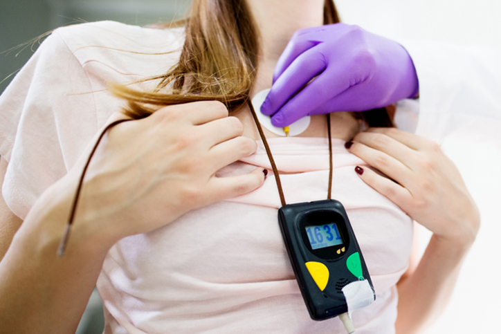
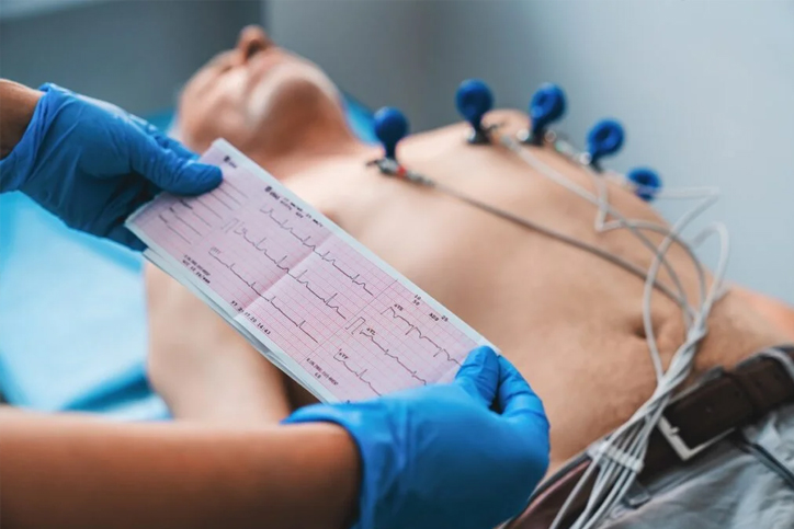
The Holter monitor is a type of portable electrocardiogram (ECG). It records the electrical activity of the heart continuously over 24 hours or longer while you are away from the doctor's office. A standard or "resting" ECG is one of the simplest and fastest tests used to evaluate the heart. Electrodes (small, plastic patches that stick to the skin) are placed at certain points on the chest and abdomen. The electrodes are connected to an ECG machine by wires. Then, the electrical activity of the heart can be measured, recorded, and printed. No electricity is sent into the body.
Natural electrical impulses coordinate contractions of the different parts of the heart. This keeps blood flowing the way it should. An ECG records these impulses to show how fast the heart is beating, the rhythm of the heart beats (steady or irregular), and the strength and timing of the electrical impulses. Changes in an ECG can be a sign of many heart-related conditions.
Spirometry is one of the most readily available and useful tests for pulmonary function. It measures the volume of air exhaled at specific time points during complete exhalation by force, which is preceded by a maximal inhalation.
The most important variables reported include total exhaled volume, known as the forced vital capacity (FVC), the volume exhaled in the first second, known as the forced expiratory volume in one second (FEV1), and their ratio (FEV1/FVC).
These results are represented on a graph as volumes and combinations of these volumes termed capacities and can be used as a diagnostic tool, as a means to monitor patients with pulmonary diseases and to improve the rate of smoking cessation according to some reports.


Medical ultrasound falls into two distinct categories: diagnostic and therapeutic.
Diagnostic ultrasound is a non-invasive diagnostic technique used to image inside the body. Ultrasound probes, called transducers, produce sound waves that have frequencies above the threshold of human hearing (above 20KHz), but most transducers in current use operate at much higher frequencies (in the megahertz (MHz) range). Most diagnostic ultrasound probes are placed on the skin. However, to optimize image quality, probes may be placed inside the body via the gastrointestinal tract, vagina, or blood vessels. In addition, ultrasound is sometimes used during surgery by placing a sterile probe into the area being operated on.
Diagnostic ultrasound can be further sub-divided into anatomical and functional ultrasound. Anatomical ultrasound produces images of internal organs or other structures. Functional ultrasound combines information such as the movement and velocity of tissue or blood, softness or hardness of tissue, and other physical characteristics, with anatomical images to create “information maps.” These maps help doctors visualize changes/differences in function within a structure or organ.
What is a CT scan?
A CT (computed tomography) scan is a type of imaging test. Like an X-ray, it shows structures inside your body. But instead of creating a flat, 2D image, a CT scan takes dozens to hundreds of images of your body. To get these images, a CT machine takes X-ray pictures as it revolves around you.
Healthcare providers use CT scans to see things that regular X-rays can't show. For example, body structures overlap on regular X-rays and many things aren't visible. A CT shows the details of each of your organs for a clearer and more precise view.
Another term for CT scan is CAT scan. CT stands for "computed tomography," while CAT stands for "computed axial tomography." But these two terms describe the same imaging test.
Hello How may I help you today?